It’s that time of year again. The time when things that should not fly take to the air. Obviously in this context it is reindeer that we are talking about but it fits in quite nicely with an article I am working on for the British Fantasy Society about other things where it is hard to explain flight.

Once you start digging into flight you realise that there are some pretty grey areas. We can explain lift in terms of a fixed wing but when we start looking at the constantly moving wings of flying creatures its gets more complicated. If we then take in the modifications that bats can make to their wing membrane it gets even more complicated. In an adaptation that was portrayed in the batman movies, but probably missed by most, bats can vary the rigidity of their wing membranes by contracting and relaxing little fibres that cover the wing surface. This can provide a rigid wing for the down stroke but a pliable membrane that can be folded for the upstroke to reduce drag.
However, let’s concentrate on something a little more festive – bird flight.
Bird’s have made a number of modifications that allow them to fly that may have escaped your notice until you start thinking about how other creatures might be able to take to the air.
Have you ever noticed how birds do not have big muscles on their backs? Muscles can only move things by contracting – they pull their two ends together. Your biceps cause your arm to bend up bringing your hand towards your shoulder but the muscles that move the bone in the opposite direction are on the opposite side of your arm. When this muscle, the tricep, contracts it straightens the arm.
Have you ever noticed that birds do not have big muscles on their backs? If you look at the shoulders of a bat they have bulky shoulders. They look a bit like a mouse who has been to the gym, but not birds.
Why don’t birds have big muscles on their backs to pull the wings back up? They have developed two very clever adaptations.
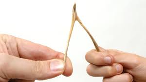
The first one is this – a wishbone or furcula. You can imagine this to be two fused collar bones. As the arms of wings come down the two ends of the bone are pulled apart. As soon as the tension is removed the bone springs back into place and pulls the wings back up. This reduces the requirement to have large muscles to pull the wings back up but doesn’t remove it completely.
Birds do have a muscle that pulls the wings back up – its called the supracoracoideus and if you have ever prepared a bird in the kitchen you have probably seen it and not appreciated what is does.
As you prepare a chicken breast you will have noticed the small fillet of meat that lies under the larger breast. This isn’t usual in anatomy. As humans we have a large pectoralis muscle and a smaller one under it and both work to bring the upper limb forwards, but that is not the case in birds

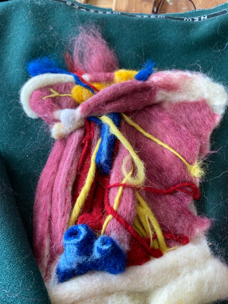
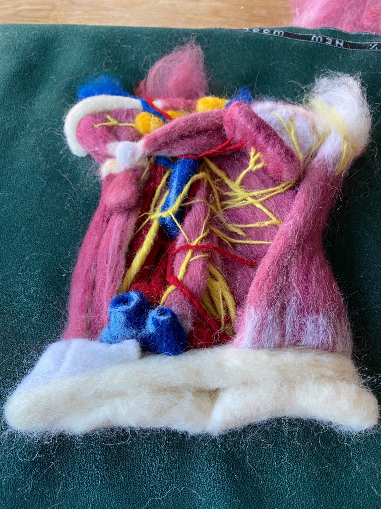
As you can see in the image. The smaller muscles found under the pectoralis major in birds is actually the supracoracoideus muscle and it actually pulls the wings back up even though it is situated on the front on the chest. It does this via a clever little looping system at the shoulder joint where it wraps around the top of the coracoid bone.
This allows birds to move their wings up and down with no big bulky muscles on the back of the bird to impinge the range of motion of the wing. It’s clever.
It does, however, mess things up if you wants a mythical creature to fly and not have a very muscular chest or to have functioning arms as well as wings.
Still, for now we can just let the angels descend to the shephards and not worry too much about how they did it.
Merry Christmas