
Last weekend saw the first National Undergraduate Neuroanatomy Competition that took place on line.
The NUNC team were absolutely on fire and produced a fantastic competition with a professionally delivered MCQ and spotter.
At the closing remarks I spotted this little felt on their desk.

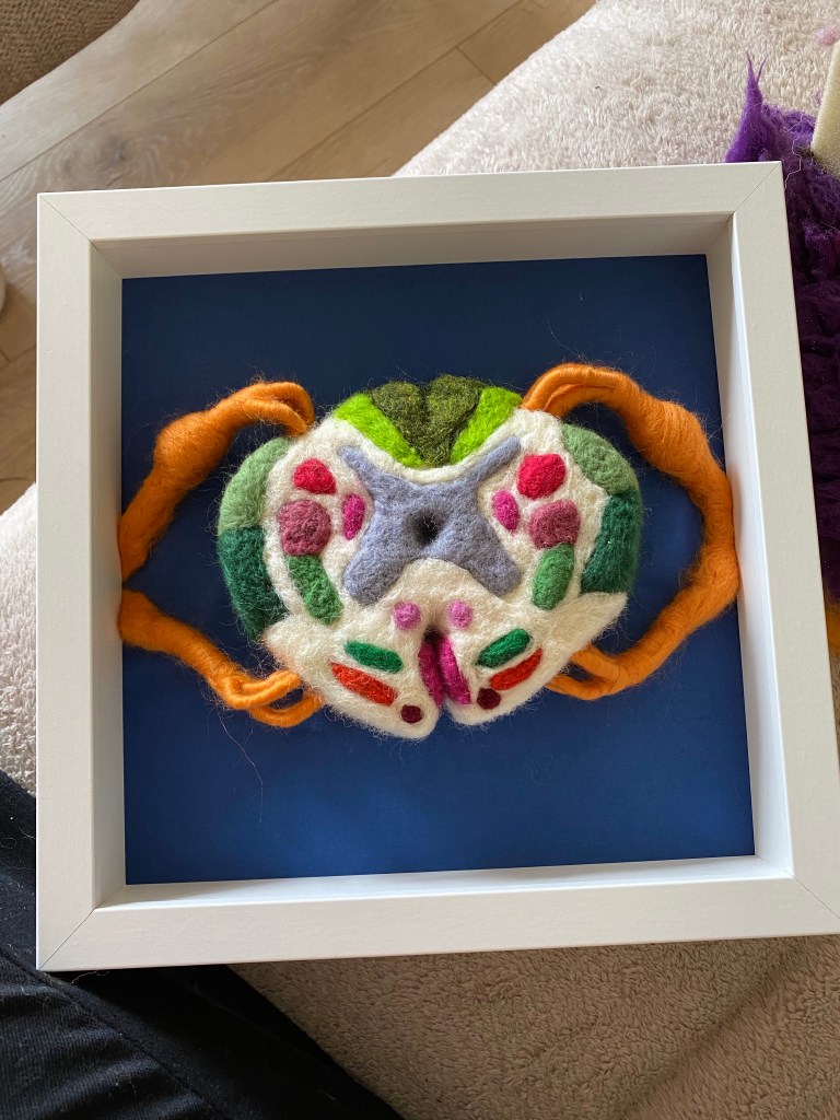
It is a cross section of a spinal cord showing the ascending and descending paths along with the nerve rootlets leaving the spine and the spinal canal in the centre. I produced it a few months before the competition and had sent it down to Dr Scott Border because he is a neuro guy (and my house is getting a bit full of felts).
There is such a thing as neurophobia; the fear of learning neuroanatomy because it is so complicated. Each of the coloured sections above carries neurons either towards the brain – green, the sensory pathways, or away from the brain – red, the motor pathways. When an injury occurs to the spine someone who understands this can work out where the damage is based on the symptoms that are displayed. It’s mind blowing.
When I learnt my neuroanatomy I studied these cross sections for ages. The information went in well enough to pass the exams.
Having spent a few hours felting the various tracts, their positions and relations to each other, it is now firmly cemented in there.
The act of doing enhances the retention of the information. Maybe learning can be combined with a craft project that helps us destress. Maybe there is more of a role for art in learning. Could be an interesting little research project.
