
There are some great medical illustrators and artists out there who produce some fantastic works of art.
Inspired by some of Sarai Llamas work, I turned to Netter to see if it would be possible to convert some of his epic dissection illustrations into felts.
The making of the neck felt is already covered in this blog on another post.
My second project would be a hand.
I started by drawing around my hand and then felting in a base of core wool to create the bones

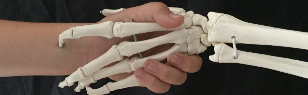
At this point I made the first mistake. I had drawn around my own hand and so I was felting a left hand whereas all of the illustrations were of a right hand.
The transition of deep to superficial is important in anatomy. It’s equally important in art. When painting you start with the sky and the background and then work forward. When felting you need to start with any structures that are going to be deep in the final felt.
The first thing to place was the radial artery.

This artery sits in what is called the ‘anatomical snuff box’ and disappears below the dorsal interosseous (between the bones) that lie between the finger and thumb.
Once this was in place the other interosseous muscles had to be placed. Each muscle runs from the length of the metacarpel (bones of the hand) towards the knuckle joint. The fibres of the wool were placed in the same direction to give the impression of the muscle fibres.
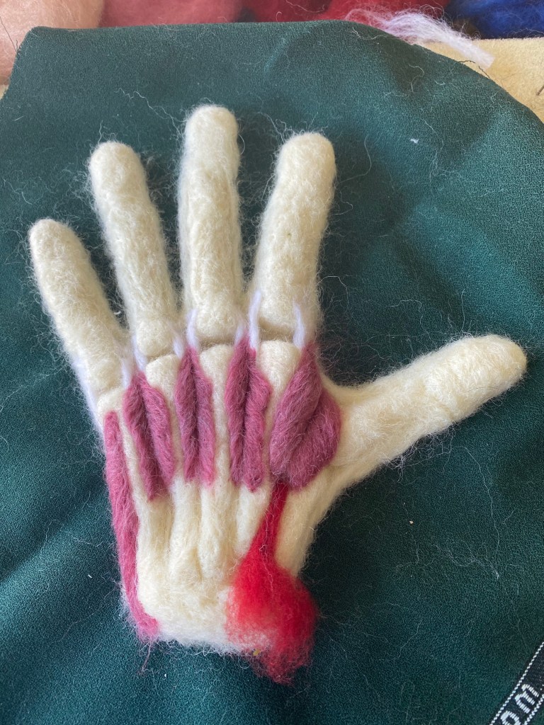
The final muscle by the little finger of the hand is the abductor digiti minimi muscle and its fibres run in the opposite direction, down towards the wrist.
At this point someone contacted me via twitter and enquired about purchasing it. I suggested they might want to wait until it was finished – it could all go wrong.

The next stage was to add in all of the tendons that connect the muscles of the forearm to the fingers. These were fine wires that were covered in wool and then felted into place.
Some of these tendons in the hand are connected and so these bridges between them had to felted into place before the tendons were fixed to the hand.
It is these bridges that give the hand some of its quirky properties.
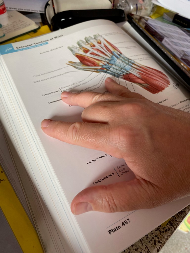
If you place you hand on a table and curl your second finger under the palm, as shown in the picture, then it’s impossible to left your ring finger up from the surface. This is due to the connections that exist between the second and third finger tendons. If the model didn’t have these connections in it then it wouldn’t depict a human hand. Ive never tried the finger trick with any other mammal so I am not sure if it’s a common feature.
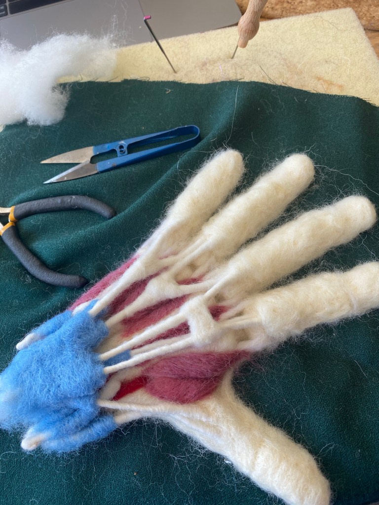
Once the tendons were in place it was a case of adding the coverings of the different compartments that group the tendons together as they pass underneath the extensor retinaculum.
The fingers were then covered in white fibres laying across the finger to represent the transverse fibres of the extensor expansions. Towards the finger tip the two lateral bands of the extensor tendon came together towards the insertion point at the base of the distal phalanx – visible in final picture.
The whole outline then had to have a layer of subcutaneous fat added in a pleasing shade of yellow before adding a layer of skin, choosing a tone that resembled my own skin as I had drawn around my hand at the start of the process.
The resulting hand was framed and packaged off to the scientist who first expressed an interest in it.
I shall return to Netter for more inspiration.

