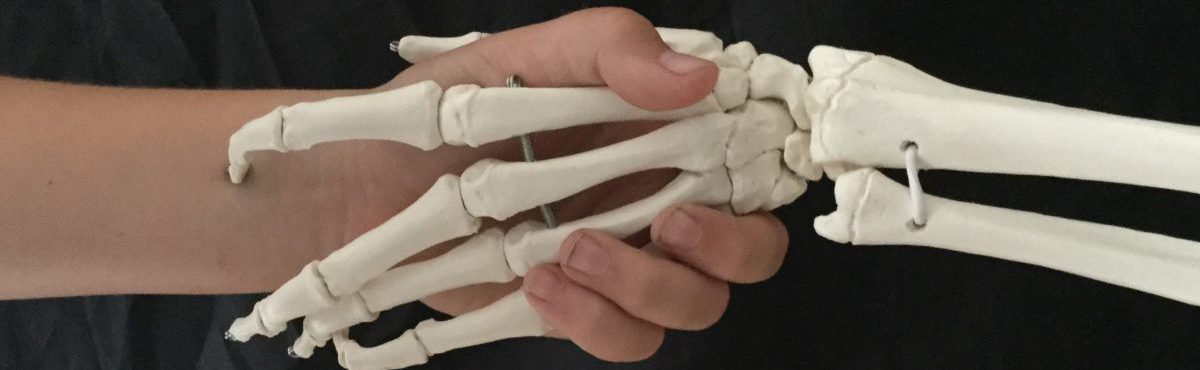
I haven’t fully divulged my past TKD experience and I haven’t disclosed I am an anatomist (why would you, these things don’t come up)
This week though my questions almost gave me away. We were looking at solar plexus strikes and I asked the question ‘and where are you saying the solar plexus is?’ I sometimes struggle with the solar plexus because its not an anatomical feature and yet, as anyone who has ever been hit in it can tell you, it does exist.
Most anatomists have assumed that when people talk about the solar plexus they are talking about the Celiac plexus – a complex combination of nerves that is made up mainly from the greater and lesser splanchnic nerves with parasympathetic innervation from both branches of the vagal nerves. It is surrounded by 10 secondary plexus so you can understand the idea that this is a central nerve plexus with radiating nerves that affect multiple parts of the body – hence the name solar with nerves radiating out.
Kudos to any non anatomists who can see the circular structure with radiating nerves in this picture!
One of the troubles that I have with the solar plexus is the mixture of worlds. If you google solar plexus then you may get one link to an anatomical site but the majority of the sites are about chakras and blocking and unblocking energy paths. I guess that is my problem with being in two social worlds that view the world differently; are you striking an energy source or an anatomical structure?
The second problem I have is that the bundle of nerves is at the back of the body just in front of the spine. To have an effect on it then you must have compressed everything in front of it. We know that can happen because we all know people who have had the wind knocked out of them – that is a sign of a blow to the solar plexus and yet we struggle with massage therapists who claim they can release the psoas by massaging the front of the body.
The body continues to pose problems for us to ponder
I recently took part in the International Feltmakers Association Felt Swap for 2022.
Every year the organisation matches up felt makers across the world and provides a theme. Each feltmaker creates something and sends it off to their partner.
This year the subject was structure.
Obviously as an anatomist there are so many things I could do about structure but I was limited by the feltmakers being mainly wet felters as opposed to needle felting which is how I produce most of my items.
I have been experimenting with a wet felted skull but it is still in its embryonic stages (ironically something else I am also experimenting with) and even with a wet felted cranium a lot of the facial bones have to be needle felted.
I decided on trying to make a model of the structure of a cell.
This is the finished structure of my cell.
The external membrane was made by wet felting around a balloon using different colours of wool to try and show the lipid bilayer.
The nucleus was my first attempt at wet felting a ball and I was very pleased with how hard the final version was until I remembered I had to get a needle through it to hold everything in place!
The nuclear envelope is essentially a small wet felted bowl. I have been trying since to recreate it in a more useful size and failing miserably.
The organelles are various wet felted shapes with the stripes of the endoplasmic reticulum (one of my favourite words mainly because my lecturer was a Geordie and it sounds even better than CurlyWurly with that accent) being a cut strip of wet felted purple wool.
The cytoplasm was my first attempt at Nuno felting. This is where wool is felted on to a thin piece of silk and the contraction of the wool as it felts pulls the material into this puckered appearance. I am thinking it might be a good way to produce a greater omentum for a project next year.
The finished project was boxed up and sent off to Switzerland where it was received with much joy.
It’s interesting to have to come up with something based on a theme and this has certainly given me lots of ideas for projects that will hopefully appear n 2023
The name Leonardo can man different things to different people.
To some it is the Renaissance polymath, to some it is a floppy haired Oscar nominated actor and to some it is a martial arts loving turtle.
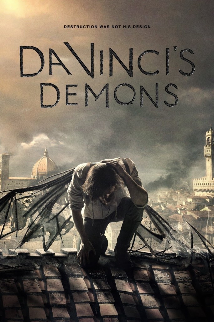
To me, he was always someone who dabbled in lots of things. The sort of things you would love to do now but unfortunately you have to pick one thing that pays the wages. This image was enhanced by the TV series Leonardo.
This image was further confirmed by binge watching DaVinci’s Demons in which he single handedly saved Florence on multiple occasions. The producers of this series claim an 85% historical accuracy but I feel this must have been focused on costume and set design rather than plot line.
Entertaining but probably not historically accurate.
Leonardo has featured in the data collection for my PhD. He is mentioned as an example of an artist who dissected. Would we allow that to happen now? Although various dramas are vague on the history, we know that Leonardo was employed as an artist. He may or may not have been employed to develop military weapons and he doesn’t appear to have claimed to have been a medic or an anatomist. He was just interested in lots of things. He was an artist who anatomised as opposed to an anatomist who drew, although I agree those two categories probably meet somewhere.
Leonardo is someone to turn to when your confidence is feeling low. A bit like Thomas Eddison, everyone remembers the things that they got right, be it the light bulb or a fantastic drawing of the human heart. Everybody forgets the thing that they got wrong; the concept that the electric chair was a good idea or his depiction of the ventricles of the brain or those tendons that support the uterus (but are found in cows not humans).
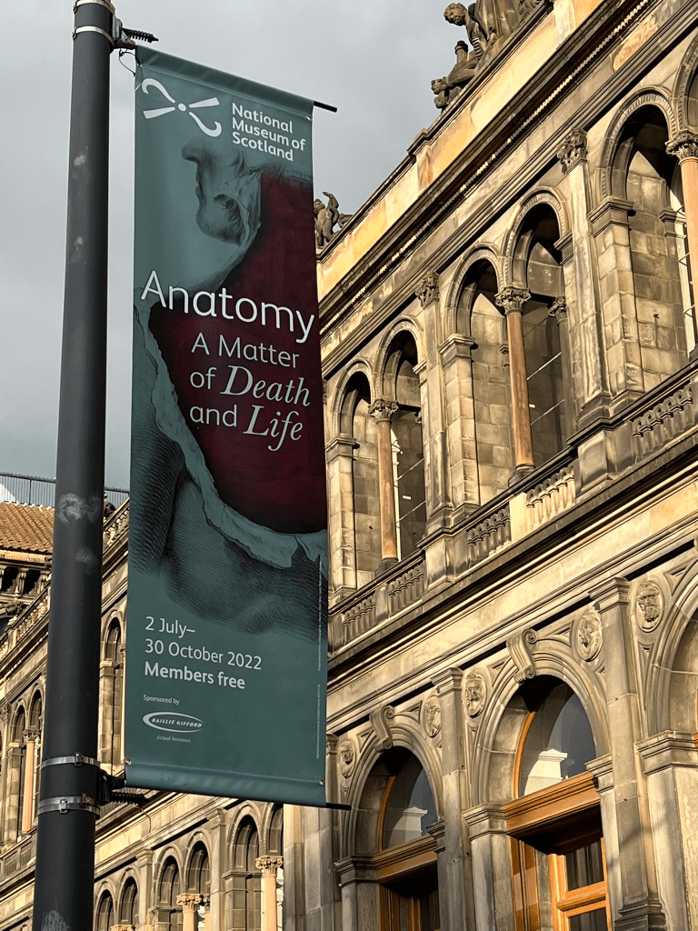
Last night I was fortunate enough to be invited along to the preview for a new exhibit at the National Museum of Scotland. This long awaited display looks at the history of anatomy teaching and dissection in Edinburgh bringing it all the way up to modern times and the need for continued body donation.
It had the potential to dwell on the gory history of Edinburgh and the body trade and yet it didn’t.
Starting with Leonardo Da Vinci – as so much seems to – the exhibitions took us through developments in Leiden and Padua with an actual copy of Versalius’ Fabrica – the first real anatomy book.
How many anatomists aren’t aware of the drawings with Clara the Rhino, and yet to see the actual picture and appreciate the size of it was amazing. Also to see the sketch on the page opposite where the arms appear to be a reasonable length as opposed to the picture with Clara where they seem very long.
There were so many images that I had seen before but had never seen the real thing and appreciated the real size. The classic picture of Hunter teaching anatomy with the model in front of the class with his arm up by Johann Zoffany, I don’t know how many times I have seen that image but to see it close up was amazing. We spent some time looking at that picture as the skeleton on the right hand side seems to have too many ribs – maybe it was a trick of the light.
The picture of Munro lit properly and behind its protective panel looked so much more impressive than where I normally see it hanging outside the staff canteen where I could walk up and actually touch it.
It was a truly cross disciplinary exhibition. Yes there were lots of anatomy pictures, a few models concentrating on the artistic aspects, but there was also a collection of costumes – robes worn by judges in the 1800s, the uniform of the town guard, there were leech pots and charms, collections of silverware belonging to clubs, complete wall sections covered with old images of Edinburgh and a mort safe (hats off to the team who installed that on the first floor).
The west port murders had to be covered – they are such an iconic part of the history but they were covered in a very unsensational way – the facts of the case, a large screen short film. It left you to ponder on what Burke and Hare were doing; killing people for medical teaching, and in a way that allows the horror of the story to sink in far better than any sensational images.
The skeleton of Burke had moved across from our museum to feature in the display but it was installed in a corner of the room, no more attention directed to it than to any other item and it completed the story and allowed you to move on.
The modern section was very well done. Three talking heads – the current Professor of Anatomy – Tom Gillingwater, a lady whose husband had donated and who was herself on the register and a 4th year medical student. They all explained the importance of dissection for education and the process of donating. The holding film between the presentation was of the names being written into the book of remembrance by the calligrapher. I wasn’t ready to see my mother in laws name been written across the screen, she donated in 2019, but I am so pleased that her donation featured in this exhibition.
The final piece was the open book of remembrance, her name again on display with all the other donors from 2019 onwards. I think she would be amused at being featured in a display at the national museum, she used to take my husband there as a child – mainly to play with the fish in the large pond in the entrance hall, but I am sure she would be honoured to be part of an exhibition that deals with anatomy and the journey of body supply in such a sensitive and informative manner.
If you can make it to see this exhibition then I would suggest you do.
Often produced as an interview challenge, the teaching of the crossing of fibres can have some people Reaching To Drink Cold Beer or trying to recall a story about two people dancing and someone else kicking them. However you chose to learn it, the complexity of explaining it to someone else can lead to confusion.
I first tried to image the brachial plexus at a glass workshop
The vertebrae of the spine are represented on the right hand side and the five nerves of the arm appear on the left. between them is the complex crossing of the nerve fibres. The limited colours belay the complexity and as a flat object it’s hard to convey anterior and posterior aspects but it worked wonders as a science communication piece. Most people think it is some attempt at a tube map and so it can be a conversation starter.
For some time I have been mulling over the idea as to how to develop this.
As a child I was in the Guides and Scouts. I was the only person I knew who had achieved the ridiculously hard knotters badge and I was frequently incorporated into camping teams as the person who could tie knots. One of my memorable moments is tying a massive sheepshank in a field with my father so that we could launch a glider that had landed and needed a shorter tow rope. For years I wondered about how to merge the macrame plant holders you see and the brachial plexus. Macrame is too complex for this – it tends to have two strands down the middle that are hidden from view – for this I needed all of the strands to be on display – I needed decorative knotting, plaiting and braiding.
This structure would be 3D – I would be able to show anterior and posterior, medial and lateral and I could use the pots to separate out the strands and hold them in place. I could have taken a knife to a plastic pot but I went a bit further and used a ‘Sculpd’ pottery kit so that I could create two pots, one with 3 holes and one with 5 holes to hold the strands apart and display the plexus.
The result is this
The 5 strands at the top represent the nerves coming from the spine. They are made up of two different colours of string (multiple strands of each) to represent the nerves that will make the medial, lateral and posterior cords – these are the three structures that pass through the first pot.
The 5 branches, musculocutaneous, axillary, median, radial and ulnar are the strands that pass through the second pot.
By using the different colours of strings it is possible to see that each final nerve is made up of components of the spinal nerves and so explain that should any spinal nerve get damaged it would not stop the nerves of the arm from working.
It took longer to figure out how to do it than to actually produce it. My house was littered with bits of string as I learnt how to braid with 2, 3, 4, 5 and 6 strands.
I’m working on writing out the instructions – I’ll add them to the web page when I get it done.
I’m also looking at the possibility of making it into a workshop so we can have more braided plexus’ out there.
Following on from my visit to the Post Mortem live event, I attended another event also entertaining and educating the public with post mortems – this one was run by Suzy Lishman CBE who has conducted hundreds, if not thousands of post mortems and the event was part of a tour to mark the 60th Anniversary of the Royal College of Pathologists. A little different from the previous event.
The two events were really chalk and cheese.
The audience was different – its hard to put your finger on what was different. This audience were maybe more professional, older, more couples rather than groups of young people. This audience looked more like the sort of people you would expect to give up a mid week evening to attend the Royal College of Surgeons, the previous audience looked like people who went to a budget hotel on the outskirts of town. (something to remember with public engagement – just because you open your doors to the public does not mean a cross section of the public walks in, sometimes you need to go to your audience)
Fifteen minutes in and it was made clear that there were no human parts being used in this event (there were a couple of museum pieces on a table behind but that was unusual for events in this tour). It wasn’t explain why, other than stating that that clearly wasn’t appropriate. Everyone seemed to agree.
They did have a model human, but in the same way that Kate Moss is a model. When the body was uncovered the person beside me commented on how real it looked just before they revealed it was actually a real person. They had apparently had someone freak out at an earlier event when the person sat up at the end and so now felt the need to tell the audience that this was a real person. This person was drawn on to show the typical incisions which were explained in detail, drawing attention to the fact that all of the incisions could be concealed so that the fact the body had gone through the post mortem process did not need to be seen. A general thread of respect that ran through this event and was missing from the previous one.
Suzy Lishman went through all of her tools, explained all the different sort of pathologists, explained she wasn’t like silent witness or Quincey and then went through what she would be looking for in each organ, the common points of failure in the human body. She spent over two hours taking us on a detailed description of what she does everyday at her work. It was fascinating. It wasn’t dressed up to be anything it wasn’t. It is what she is trained to do and does for a living.
She talked about the texture of the brain, the slicing of every organ, the reason they don’t cut the top of the head straight off but leave a little upturn at the back. She could have made it sensational – but she didn’t – because it’s not appropriate.
In the questions at the end she was asked whether she had seen any surprising things. She said she had seen some truly amazing things but they were so amazing it might be possible to identify the person and therefore she wouldn’t be talking about them.
The thing that struck me is that this is a person who deals with dead bodies for a living. They have an inbuilt sense of what is appropriate and what is not. They can still make the human body fascinating because their own passion shines through. There is no need to dress it up as something it is not or to sensationalise it in any way. The body is amazing and we should appreciate it for what it is.
At a fifth of the price of the other post mortem event this is where you should go if you want to learn about post mortems. If you want to dissect organs then there are other events for that that make it clear they are using by products of the animal food industry.
Today is the installation day for a new exhibition in the Anatomical Museum at Edinburgh University looking at Colonising Mars. Our team looked at anatomical changes.
Here is our piece – Life on Mars with David.
Let me start by acknowledging that David was carved as a white man and we are meant to be decolonising everything but we went with David – despite the known issues about proportions and it being carved so that it looked correct when viewed from below – because it was easier to show a colour change and we had a pop culture reference to address.
Also acknowledging the words of the evolutionary anatomist we consulted that ‘trying to predict evolution changes is an exercise in futility’ – view the piece as a conversation starter.
Also – selection pressure isn’t really a thing anymore as we do not allow the weakest in society to die (quite rightly). It is no longer an evolutionary advantage to have certain physical traits.
All that been said – What did we change?
Height – The gravity on Mars is 38% of that on Earth. It is the resistance to gravity that gives our bones their strength. Bones lose up to 5% of their mineral density per month of space flight. The reduced gravity would lead to more fragile bones and hence increase the likelihood of fractures. We thought we would address this by developing a stockier frame to protect against fractures.
Head size – as bone fractures are more likely we thought that birth would be more likely to proceed by C section. There is a debate that it is the passing through the birth canal that limits the size of the human head. With this restriction gone, head size could increase. (Mitteroecker, P. et al (2016) cliff edge model of obstetric selection in humans PNAS,113(51))
Eye size – the light on Mars is the equivalent to a cloudy day on Earth. The adaptions that the eyes might make would probably all be internal but that doesn’t work on a model, so we made the eyes bigger.
Skin – the annual exposure to radiation is greater on Mars – 30 millisieverts as opposed to 3. For some reason all of the people discussing colonising mars have increased the pigmentation in the skin with carotenoids rather than melanin. We couldn’t find out why but increased the orange colour to fit in with the prediction that skin pigmentation would increase.
Spleen – we developed a bulge on the left to indicate a larger spleen. We based this on research on the Bajau people who spend a lot of their time free diving. These people have larger spleens. Upon exposure to an oxygen restricted environment- part of the divers response, the spleen pushes a large volume of red blood cells into the circulation so that the breath can be held for longer. We thought there would inevitably be some sort of breach of the containment on Mars and so the ability to hold the breath for a longer time might be helpful and might actually be a genuine evolutionary selection criteria. (Trenkman,M. (2018) A breath holding adaptation. Nature Review Genetics 19.)
Limb circumference – some of our body shape is due to the distribution of fluids within our tissues. This is affected by gravity and so a reduced gravity would redistribute our fluids. According to NASA the circumference of the leg can reduce by 30% during space flight (sitn.hms.hardvard.edu/flash/2013/space-human-body/). We wanted to reflect this effect and so reduced limb circumference and removed the chiseled abs.
We hope it starts a conversation and maybe makes people aware of what parts of their bodies’ do and why they are like they are.
This is a long post with only the one picture for reasons that will become apparent
This weekend I attended the Post-Mortem live event. It was a hard decision, but it is talked about a lot in my research and some people who have never seen it have views about it. In the same stretched logic that led me to read the infamous 50 shades of grey before voicing an opinion, I thought I should go and see it.
(It should be noted that I was not neutral about the event before seeing it. I am an anatomist. I did my training in the dissection rooms of UK medical schools. I am not a pathologist. I have never attended a post-mortem and my experience in that area is confined to Dame Prof Sue Black’s books, Carla Valentine documentaries and TV. I am not an entomologist, I have not studied forensics and I have not conducted mass spec or gas chromatography. I very much doubt any one person has.)
The event was 4 hours long, although the doors did not open until it was meant to start, and it had a 35 minute break so it was closer to 3 hours.
I help run the anatomy night events where we run dissections which are usually just over an hour. In that time the anatomists cover one organ. We are currently submitting a manuscript on the educational value of those events.
This event covered crime scene analysis – 2 separate potential crime scenes, collection of swabs, kidney dissection, soil analysis, DNA analysis including the explanation of exons, introns and small tandem repeats, soil analysis, entomological study of fly larvae to estimate time of death, viewing the body under UV light to identify sperm samples, craniotomy and removal of the brain, dissection of the larynx, bronchi and lungs, and heart.
To be fair, in the one page programme which the majority of the audience did not appear to have – it cost extra, there were QR codes to all of these techniques and a paragraph that said that the show depicted something that was very far from the normal post mortem process but this had been driven by feedback from2021 where guests said they wanted more grit and forensics and to find out what happened.
The issue with this event is the pretend use of human remains. In this case a body sitting at the front of the stage that had supposedly been dead for around 16 days. You might think – who would be fooled by a synthetic model, but the vast majority of the population have not seen a dead body – they have no comparator and when the remains are there with pig intestines added to the abdomen you could see some people thinking this was real.
In the queue outside, the person behind me talked about the fact that he was a registered body donor. ‘This could be me in a few year’s time – still it’s the learning that is important.’ He said he was surprised the event wasn’t taking place in a hospital or a university. He did approach the presenter during one of the activates and I did overhear them say that it was not a real body as that would be illegal, but no room wide announcement was given. The potential donor standing a few feet from the model had to be told it wasn’t real. I can’t see anything in the programme that explains the origins of the body.
There were a number of people in the room who were there for the same reason as me. There were several students who were there because this was their only access to dissection. There were some people there who were thinking of going into forensics (this misrepresentation of the role of a single forensics person feeds the forensics frenzy), some thinking of going into medicine and a bunch who were just interested. The audience covered a wide age range but was probably more towards the 20-30 year age group. There were a total of 112 people with tickets costing around £50-£80.
The presenter had spent 5 years studying anatomy and had worked at the Royal College of Surgeons. (I had previously tried to find out the qualifications of the staff at these events and was told by the company that it could not be disclosed because of data protection). Their anatomy was only challenged by the limits of the show. They cut the cranium off with a scalpel, they mentioned a bone saw but obviously didn’t use it. They removed the cranium and the model brain literally fell out – it wasn’t connected to anything. When it came to the digestive tract – which was stained blue due to an attempted poisoning, they had to explain the lack of an appendix – why on earth they didn’t just say the victim had had an appendectomy I don’t know – I guess saying the same thing twice a day for several months was wearing a bit thin, but they suggested that the appendix often just falls off or explodes.
Except for the two points above, there didn’t appear to be anything wrong with the anatomy. I can’t comment on the fly larvae each group had to go up and collect from the corpse to work out the timeline and sticking a pH metre into the soil sample to differentiate between the two crime scenes was a time filler. Even having qualifications in medical microbiology and anatomy I found the speed at which they went from ‘this is a kidney’ to loops of henle, to mass spectrometry to DNA profiles startling and some of the slides of the power point were flashed up so quickly it was impossible to read then. The room was dimly lit with spot lights at the front facing the audience which made it hard to see although they had no issues with people crowding around their pretend human remains and taking pictures. They had a camera operator who zoomed in on certain thing that were projected on to two big screens.
Each table had a bucket and a tray and got to dissect a kidney, identify fake larvae, open by a larynx, trachea and lungs and a heart as well as take the pH of a soil sample. Several swabs were taken and sent off to a lab with results coming back during the event. Dissection instructions were given verbally, and we were provided with scissors. The one presenter toured the room to check that people were getting on OK. The trachea had had a black substance (possibly charcoal solution) placed in it to suggest particulate matter that had been breathed in.
If the event has been called who killed Miss Piggy then I would not have any problems with it. It would have been quite entertaining although maybe not as marketable. The event could even have run without the pretend rotting corpse at the front of the room. I would have liked a big sign up telling everyone that they were dissecting pigs remains. Some of the audience even commented on the amount of pig remains that were being used and speculated that they were probably been thrown away afterwards – maybe they were going back to dog food – I don’t know.
I left fairly unimpressed. Unimpressed with human kind in general that we find the idea of paying to look at dead bodies acceptable, and unimpressed that there are business people out there prepared to exploit it. The programme claims that the company supplies over 40 UK universities and a dozen NHS trusts although this applies to the ITAE event management and production company, maybe not the post mortem section of the company. Disappointed that there are body donors out there who think this is what might happen to their bodies and also slightly worried that it didn’t actually seem to concern them.
Next month I am booked into the Living Autopsy. An event being run by Dr Suzy Lishman CBE as part of the 60th anniversary of the Royal College of Pathologists. It will be interesting to see the difference.
You can’t get around depictions of Anatomy – as soon as you draw anything you have depicted anatomy and unless you are adopt a painting style that is some what abstract, you open yourself up to claims that your depiction isn’t accurate.
A recent literature review uncovered several publications where modern anatomists were pointing out the errors of famous artists – a future blog post maybe.
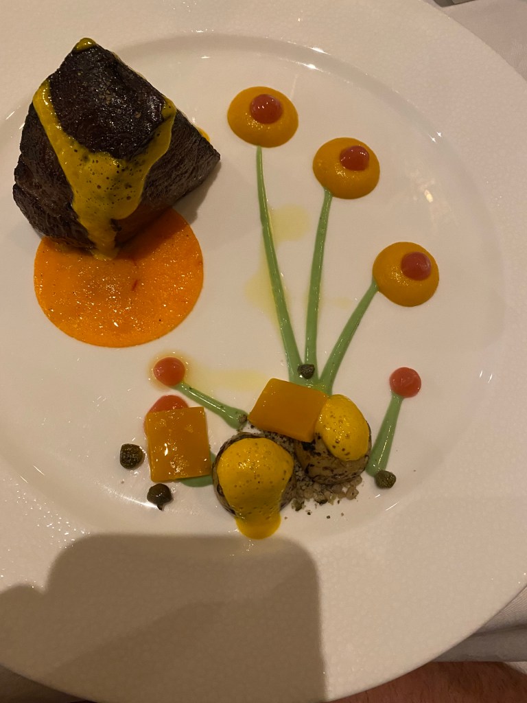
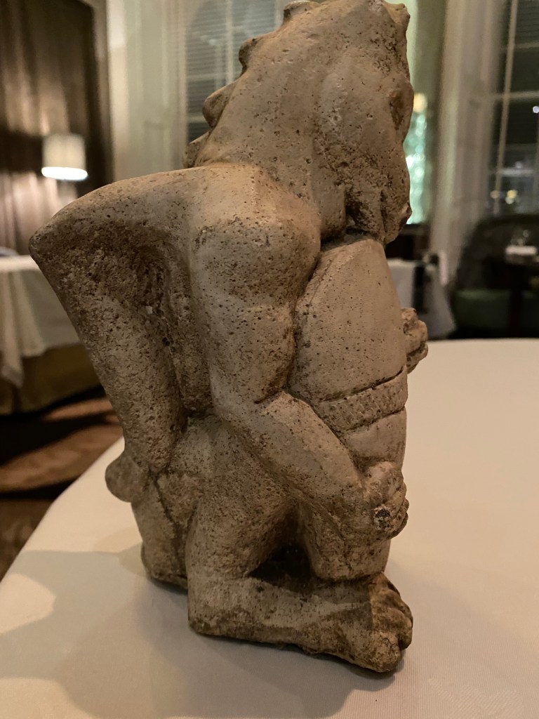
Last week I was out for dinner at the Edinburgh restaurant 21212. It had been on our list of restaurants to visit for some time. The food was fantastic, and the presentation was outstanding.
Each table had a small figure on it. The table beside ours had this little model of a dragon.
I had spent some of the previous week finishing an article for the British Fantasy Society about winged mythical beasts and the problems they present for anatomy.
You can see that this dragon has wings and shoulders which poses questions as to how either limb works without effecting the other. There is always something around to make start a conversation on anatomy – this is a good one as most people don’t fully understand the relationship between their arm and their shoulder blade and when you add breathing in to that equation they rarely appreciate the fact that they are all connected.
Upstairs, in the private dining room, there is an amazing picture
This is a small part of the painting by Caravaggio called the 7 acts of mercy. The seven acts are actually all depicted in the lower part of the painting and this section just shows two angels looking down on a scene that we can no longer see.
What we can see though are pairs of wings coming out of the back of the angels. How would that work? An excellent backdrop for a science talk about anatomy of flight – somehow I can’t see the science fair budget running to a private dining experience.
In 2016 I wrote a book on William Burke, the provider of bodies for medical teaching in 1828. I am based in Edinburgh where tales of Burke and Hare are everywhere and tourists have easy access to various versions of the truth.
That was the main driver to get the research done and get as close to the real story out there for people to read.
Since 2016 I have been speaking at various events, turning up on TV screens and been in receipt of various documents that continue the story of Burke and Hare.
Late in 2021 I discovered that the cell that held Burke after his conviction was still in existence. Its not open to the public but it is preserved under the courts in Edinburgh.
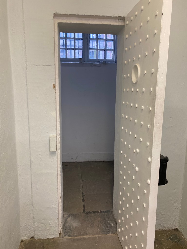
I was lucky enough to go down there – it is tiny. We know that Burke complained that he couldn’t see the sky from his cell because of the height of the buildings behind the court. The window is 75 inches from the floor, so quite high up anyway and if the sky couldn’t be seen then there wouldn’t be a lot of light getting in there. The door is solid metal with a tiny peep hole. The cell would have been in almost complete darkness. The room itself is 63 inches by 61 inches. We know from the skeleton that Burke was 5 foot four so it would have been impossible for him to lay down in the cell apart from on the diagonal and you have to assume there would have been some sort of furniture in there if even just a cot and some straw. Obviously prisoner comfort was not a priority in those days but I’d like you to take a moment to compare it with the cells other people were held in – pictured below and now full of cabinets.
Burke’s Cell
Other cells
When I asked why there was such a difference, my guide suggested that they just didn’t like what Burke had been up to and they were just being mean. He lived in that cell for 1 month.
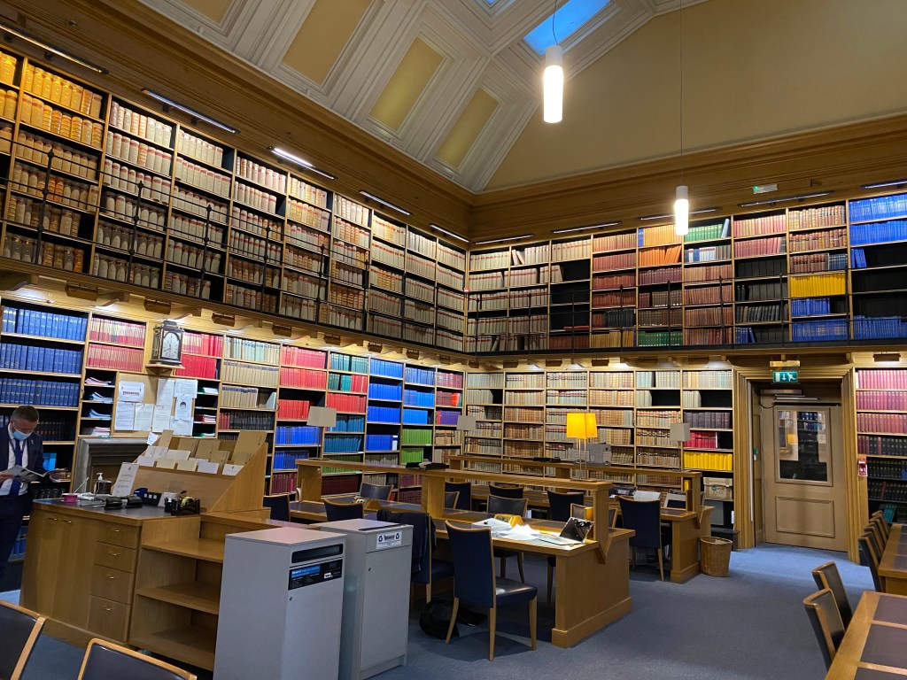
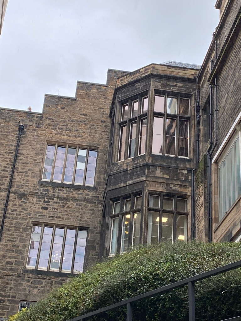
The court where the case was heard is also still around, now a library for advocates. The second picture shows the windows of the court room from the cowgate. Not quite sure how a crowd gathered under these windows to hear the court case – I may need to go back and look at plans to understand if there were more windows.