I had to share this fantastic piece of work by Chris Rynn who has brought the cover of my book on William Burke to life.
Chris performed the original facial reconstruction of William Burke for my Book cover about the murderers who supplied bodies for anatomical dissection in the 1820’s in Edinburgh.
The reconstruction was based on the skull which is in the Anatomical Museum of the University of Edinburgh and also the life and death masks which are also held in the museum.
The new AI that was released last week has allowed a sense of life to be given to the reconstruction.
If you want to find out more about Burke you can get hold of the book on the shop page here or on amazon.
I suspect we all know by now that Tiger Woods has hurt his leg in a car accident.
We can all wonder at the fact that an accident which a decade ago might have resulted in amputation can now be fixed with metal rods and pins.
What a wondrous opportunity to engage the public in understanding a little more about their bodies.
This was the picture from the British press.
TIBULA!
He’s broken his Tibula!
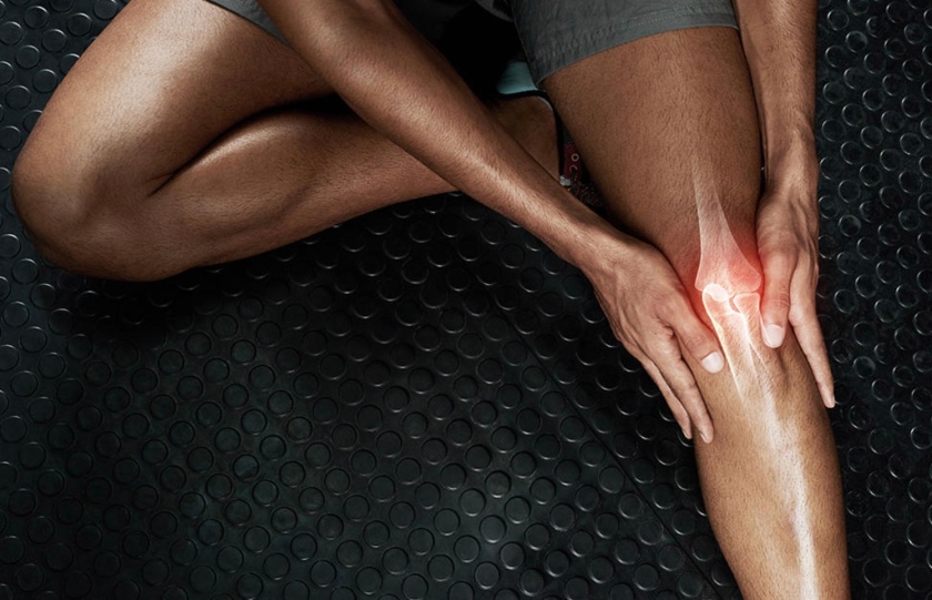
I suppose we shouldn’t be too harsh at least there are two bones in there and they are the correct bones not like this unfortunate runner who is suffering from knee pain, mainly because that is an elbow joint in his leg.
The main bone in the leg is the Tibia. Before you correct me that it is the femur, remember there is a difference between the thigh and the leg.
Tibula was an ancient town in Sardinia according to Goggle but actually the same google search brings up several pictures of legs, an article on orthopaedic surgery and getty images, where a search for tibula will bring up clip art and stock images of leg bones.
Maybe as anatomists we ought to be out there being more vocal and acquainting people with the parts of their bodies.
It’s a wide open dish that is used to hold vinegar. This particular one came with a set of flavoured vinegars. Its open and wide so that you can dip your bread into it.
It may seem a strange object to link to anatomy but this highlights one of the things I love about anatomy – the language.
A lot of the terms in anatomy seem very complicated; muscles have big long names, and yet once you ‘crack the code’ those names tell you so much and there is not a single word wasted.
Is it long and straight? – it’s rectus. Does it contain the word superior? Then it will be closer to the head than a similarly named structure that will be called ‘inferior’. Is it anterior? Then there will be one behind it called posterior. Does it contain the phrase ‘levatator’? Then it will lift something.
Once you have cracked the code then you can guess the name of structure by knowing what it does and vice versa.
How does this relate to a dish for vinegar?
There are some structures in the body that are named after structures they look like.
The structure in the middle of this picture is an acetabulum.
It is the socket in the pelvis that the femur (thigh bone) sits in. Hopefully you can see the similarities between the two structures.
Anatomy is littered with these helpful cues that can be used to remember the names of structures. You could refer to the joint above as the hip socket – it wouldn’t remind you of the shape. Would it be easier? – maybe.
I’ve recently discovered that some people are taught about the pelvis in a way that I’m sure their instructors think is helpful. I’m not convinced. Join me in the next post where I’m going to look at the bony pelvis and some of the structures around it.
The Christmas break gave me the opportunity to finish off a prize for another anatomy related competition.
This skull full of flowers is being offered to the winner of an image competition currently being run by Anatomy Nights.
It is open to anyone, please go and check out the details here. Please let everybody know about the idea and help us flood twitter with anatomically inspired images in February.
I’m hoping 2021 is going to enable me to bring some more content about anatomy that is going to add to your day and your enjoyment of life.
Then I’ll begin. 2020 has been a strange year for many of us. A lot of us have lost the commute to work and we are spending longer than ever sitting in front of a screen, often in places that were not designed for office work.
The slouched head forward posture of the office
We were not great at how we sat in chairs. We slouched and adopted a head forward posture. Now we have to contend with people trying to work with chairs that were not designed for office work, some even forced to use the sofa or the bed. It has been a challenge.
A few weeks ago I had someone complain to me about their sacrum being sore through all this sitting. I didn’t say anything at the time but the comment kept chipping away at the back of my brain.
You shouldn’t be sitting on your sacrum.
Your sacrum is a wedge shaped bone at the back of your pelvis, so called because it used to be believed that it was the sacred part of the body that if planted could grow into another body (!). It is often described as a key stone structure that sits between the two halves of the pelvis and that gravity acts on it to wedge the two halves of the pelvis apart. A belief that is clearly not the case if you look at the position of the pelvis from the side where it can be seen that the sacrum is more like the roof of the pelvic cavity that the back wall – I digress.
It can be seen from this image though, that if you were sitting then you shouldn’t be on your sacrum. So what should you be sitting on?
Your pelvis is made up of three bones, The ilium, the pubis and the ischium. (It is a long standing point of disagreement as to whether the sacrum is part of the pelvis or not). These three bones fuse in childhood to give us the distinctive hip, or pelvic, bone. At the bottom of the pelvis, on the ishium, we find a bone lump which anatomists call a tuberosity. These tuberosities are called your ischial tuberosities, because they are on the ishium. The exercise industry has decided to make it all a lot simpler and refers to these structures as your sit bones – because you sit on them.
There are a number of muscles that attach to your ischial tuberosities. If you sit on a hard surface and rock backwards and forwards you may be able to sense when you are on your sit bones but if you have a reasonable amount of body fat (and we all need body fat) it may be a challenge to actually palpate them.
view looking upwards between the legs, front of pelvis at top and sacrum at the bottom
The ischial tuberosities divide the area between your legs into two triangles which anatomists refer to as the urogenital triangle, because it contains the openings of the urinary system and the genital opening, and the anal triangle because … it’s self explanatory.
When you are sitting on your chair you ought to be balanced on your ischial tuberosities thinking about your weight going down through the urogenital triangle rather than the anal triangle. As you sit like this your head will come back into a position over your neck and you ought to get less strain on your neck and shoulders and most of all … no pressure on your sacrum.
Let’s hope we will soon all get back to our ergonomically designed chairs and we can sort out all the aches and pains brought on by home working.
In this time of lock down I was lucky enough to get away for a few days at the end of August. We went down to the Lake District in England.
To get there you have to pass along this beautiful valley called the Devil’s Beef Tub. The road south from Edinburgh to Moffat runs along side the valley. It twists and turns, passing over bridges and streams that tumble into little waterfalls.
It is at one of these turns and picturesque stone bridges that we can relate it to a mile stone case in forensics and anatomy.
Bella Ruxton, the wife of Buck Ruxton
In 1935 a Lancashire doctor Buck Ruxton took it into his head to murder his wife, Bella, and her maid Mary. Finding himself with two bodies to dispose of he decided to dismember them and dispose of them in the Devil’s Beef Tub. He wrapped them in a local Lancashire newspaper, a fact that would come back to haunt him later, and scattered the 70 pieces of remains into the countryside. Some of them were found by the local police and one of the most famous murder investigations started.
In 1935 forensics was still very much in its infancy. A super team of experts was formed with Glaister, the Glasgow professor of forensics, Smith, the Edinburgh professor of forensics and Brash, the Edinburgh professor of anatomy, leading the way. They very soon reached the conclusion that the bodies had been dismembered by someone who knew what they were doing. Building the grizzly jigsaw soon informed then that they were looking at the remains of two individuals but they still had no idea who, or even what sex the victims were. When they were identified as women the penny dropped that maybe this was related to the two women who had gone missing from Lancashire, reported missing by Buck Ruxton, a man who had the capability to dismember.
The finger tips has been removed from one victim hoping that this would remove the ability to take fingerprints. The other victims hands had decomposed beyond the point of being able to finger print the skin. The newly formed finger print unit in the UK worked with the FBI to take fingerprints from the dermal layers that were left on the hands and managed to produce prints that matched in over 16 positions to those of Mary the maid which they had managed to lift from the house in Lancashire.
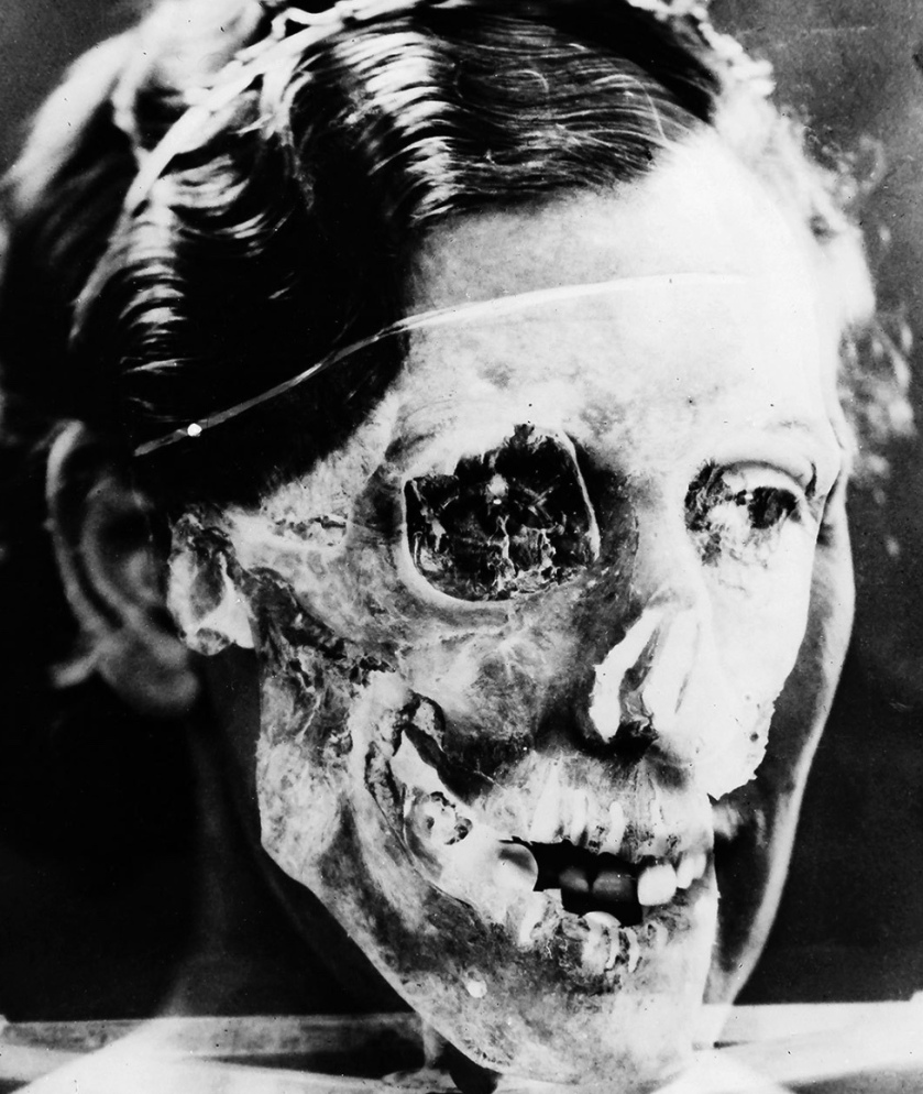
This left the puzzle of how to positively identify the other remains. The skull had been recovered but they were struggling to get any identifying features. Then one of the team came up with an idea. At that point it was ground breaking. Now, it is a common feature of masters students projects. Could a picture of the skull be superimposed onto the portrait of Bella. She had a very distinctive jaw line; could it persuade a jury that the remains were her’s?
After much positioning of the skull to match the image above the team were able to produce the picture below
As you can see the ear, the eye socket and the jaw line all match.
It was proof enough for the jury who decided that the remains were those of Bella and her maid Mary.
Buck Ruxton was executed in Strangeways prison in 1936 and the case went down as the first modern murder investigation.
Full details can be found in the book by Tom Wood, Ruxton – the first modern murder but bear in mind it might change your view of that beautiful valley if you travel down that road.
Applying science to reading fiction can be likened to golf; it can be a good walk spoiled. Whilst I groaned when the original Bladerunner allowed someone to see something that just wouldn’t have been possible in a mirror, I will quite happily accept a fictional dragon.
I’m reminded of a comment I heard last week – gravity always acts downwards. Somethings in science should not be altered. What you can see in a mirror is governed by the laws of physics – it should always be the same. How a dragon flies is not governed by science. They don’t exist in reality. We don’t have anything with wings and forelimbs because our knowledge of anatomy would not allow it. It can be quite arrogant to say that because we don’t understand it, it can’t exist. It’s certainly not an attitude that is going to take you very far in reading science fiction.
We can use it the other way though.
One of the key ideas with Science Communication is meeting the people where they are. They have an understanding of science fiction – can we use it to engage them in science?
I had my sci fi aha moment when my son was studying higher biology. Well, he was meant to be but he was actually spending most of his time watching the Walking Dead. Over breakfast one morning we started a conversation over which part of the brain would have to be affected to make the zombies lose the ability to speak. It grew from there. An hour later we were still talking about neuroanatomy and what he had studied at school. That summer I appeared at the local Science festival talking about the neuroanatomy of zombies.
Last weekend it went international when I joined the panel at Gen Con to talk about the science of science fiction. Indianapolis science had two panels. One looking at the science behind mythical beasts – is Chewbacca more like a primate or a dog? and the other looking at zombies. I’ve attached the links to the you tube films below. It was great fun joining in with an event like that – even if the time difference meant I had to stay up in to the early hours of the morning!
Science shouldn’t be used to destroy the joy of science fiction but science fiction can be used to enhance science. I’m aware of one medical school that has a session in their anatomy course where students have to design an animal made up of at least three others. It might seem a little strange but just think for a moment. If you are going to combine three animals then you need to understand how all the vital systems within those animals work in order to combine them. If you want to take part in a discussion as to why we don’t have creatures with forelimbs and wings then you need to understand the musculature of forelimbs and wings.
Maybe we can move forward together.
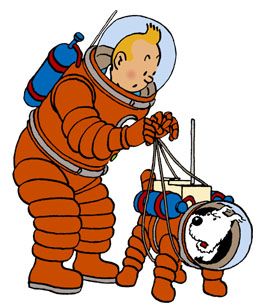
The presence of science in fiction first came to my attention in Tintin. In Destination Moon Tintin goes up into space. In the comic they show the balls that water forms in zero gravity. A great use of science in fiction…it was written in 1950. That is 11 years before the first man went into space. A great example of the fact that the author doesn’t always know what they are writing about and science can’t always be used to explain it.
Links to Gen Con Science of Science fiction panels
As Anatomists, we should be used to things being named in a way that is not helpful or often misleading. The world of eponyms has left us with literally hundreds of structures in the human body that do not provide us with any information other than who first claimed to identify it, the great vein of Galen, the Fallopian tubes and the angle of Louis to name a few.
It should not surprise us to find the same issues in other professions. Let me introduce you to the figure four choke hold in competition grappling.
There is the first issue of what is the difference between choking and strangling. This is as productive and circular as to whether jam or cream goes onto a scone first. I go with the simple, choking is an internal issue – you can choke on a piece of chicken, whereas a chicken has never strangled anyone. This creates the first issue for the pedant. The uninitiated pedant once experiencing a figure four hold might think ‘this opponent doesn’t know what they are doing. There is a large gap at the front of my throat. This isn’t a choke hold.’ and they will continue to think that right up until the moment they pass out, because the choke hold isn’t designed to choke you.
There are a lot of important structures in the neck. There is the airway or trachea. This is held open with C shaped pieces of cartilage so that it does not become compressed as the head moves around. We have the oesophagus which connects the mouth to the stomach. This is a stretchy muscular tube that spends most of its time closed anyway. We also have a lot of blood vessels as you can see in the picture. The two most important are the carotid artery and the jugular vein. They run up each side of the neck taking blood to and from the brain. They can just be seen appearing at the top of the sternocleidomastoid muscle running from the back of the head to the front of the chest (from the sternum to the mastoid process – an example of how things should be named).
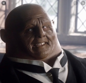
If we had necks like the Sontaran from Dr Who then this wouldn’t be an issue but we have accepted the vulnerability of exposing all of these structures in a long slender neck for the ability to turn our head around and be aware of what is going on around us.
The figure 4 hold works by compressing these vessels, restricting blood flow to the brain and causing people to become unconscious. That is where the discussion should have finished but…a little knowledge can be a dangerous thing and we now have two other debates appearing.
Is it the compression of the jugular vein or the carotid artery that causes the unconsciousness? Veins are quite flat and easy to squash whereas arteries have springy walls and are better at holding their shape. The jugular vein is more superficial than the artery so it probably gets squashed first. I would be very suspicious of anyone who claims they can compress one and not the other. The figure 4 hold is a blunt instrument to an end.
Even more ludicrous then is the discussion around the carotid body. The carotid body is a small structure that sits just above the bifurcation of the carotid artery. It has a role in controlling blood pressure through out the body. Theoretically if the carotid artery were compressed below the carotid body then it would perceive a drop in blood pressure and initiate the process to increase the blood pressure. If the artery were compressed above the carotid body, it would perceive an increase in blood pressure and initiate steps to reduce it. The outcome in terms on consciousness for the opponent would be the same in both circumstances. The carotid body is about the size of a pea and, due to anatomical variation, not always in exactly the same place in each person. The pressure is being applied with something varying in size between a forearm and a thigh. I don’t think I need to point out the ridiculousness of claiming any precision in its application.
I was recently asked why arm bar’s are so effective. These questions usually come from people who have not experienced arm bars – one of the most effective submissions moves in grappling. This time however, I thought about the anatomy of it and the effect this might have had on the exercise world.
An arm bar, correctly applied, usually causes a tap out within a few moments. If you can imagine lying on the ground. Your opponents legs are across your chest and neck, effectively pinning you to the ground. Your arm is extended between their leg and your hand held palm up on their chest. This might already be causing you some physical discomfort depending on the flexibility of your shoulder and the size of your opponent. Now they start to push their hips up. The discomfort escalates as your elbow is forced to hyper extend. You are not getting out of it and most people tap out.
Why does it hurt? Just look at the connective tissue around your elbow.
Every ligament and tendon is filled with receptors that you usually use to help locate them in space. They constantly feed back information to your brain that tells it how much stress it is feeling or how quickly it is accelerating. The brain can calculate which direction things are moving in by comparing the messages from different parts of the body. And it does all of this without you even thinking about it.
Have you ever fallen asleep when you shouldn’t have and woken up with your head jerking back? You probably thought you work up and jerked your head up to appear awake. It happens the other way around. When you fall asleep your head will tip forwards as the muscles relax, due to its weight. This causes the muscles and connective tissue at the back of the neck to suddenly be stretched. That sudden stretching sends a message to the brain ‘We are stretching too quickly, slow it down.’ The body slows that down by contracting the muscles that oppose that stretch. This causes the head to jerk back up and that it what wakes you up. Your body took care of its self whilst you were asleep.
As the muscles and connective tissue start to get stretched in the arm bar the brain gets the same messages. ‘We are being too stretched’ Only the arm is trapped, it can’t do anything to reduce that stretch.
Two bones make up the forearm; the radius and the ulna. The ulna has a large hook like prominence that makes up the point of the elbow. When you straighten your arm, this prominence fits into a hollow at the back of the humerus, the bone of the upper arm. If you continue to extend it then the bone can go no further, it is blocked up the humerus. I’m told it is very unusual to break this prominence off of the ulna by using an arm bar – the usual injury is a dislocated elbow. Either way it is painful.
But what does this have to do with the exercise world?
Have you every gone to a legs, bums and tums class. Endless squats but usually also endless bridges. You lie on the ground, feet close to your butt, and push your hips up as high as you can. A great exercise for the glutes but where do you think it came from? The origins of the bridge can be traced back to ancient Greece and wrestling. The easiest way to get off someone who is on top of you is to push up your hips. The best way to get a submission is to apply an arm bar. The best arm bar will be applied with the highest hips. We might use the exercise now to get a shapely bum but that isn’t why it was developed.
Janet has a PGDip in Anatomical sciences and is currently studying for a PhD looking at different groups beliefs about the body. She represented the UK in TKD and learnt grappling as part of her Budokon belt system.
By the end of May, two months into lockdown, new forms of entertainment were needed. Into this arena appeared a company who had made their mark on Dragon’s Den two years previously. They were offering a live post mortem!
I had heard of these events before. They usually tour the country offering dinner and dissection for the same price as a Michelin starred meal. The events usually sell out. In the name of research I watched the recordings of post mortem live.
They use a semi synthetic cadaver. The question of what exactly this was appeared on twitter and was not answered. It appears to be a synthetic human shaped shell into which they place pig intestines for the performance. It was covered in plastic for almost all of the performance causing some people to complain about the glare. I expect it was expensive and they wanted to protect it. If you read a long way down on their web site you do find the mention of pig but there was not one mention of it during the 7 hours of broadcast. In fact they seemed to go out of their way to imply that the figure was human. Several people made comments about the human specimen on twitter which were never corrected and several people even thanked the donor.
The presenters, we were told, were anatomists of national acclaim. I’m not sure what that means. One of them was referred to as Dr xxx PhD. This is confusing in the UK. Is that a medic with a doctorate? He introduced himself as a final year medical student (equally confusing in a year when they accelerated the graduation of final year medics to deal with the pandemic) so the Dr and the PhD are actually the same qualification. The company refuses to release the qualifications of its staff, saying it breaches GDPR.
There was nothing wrong with the anatomy. A few mistakes, but who doesn’t make mistakes in a live broadcast. The ease with which the organs were removed from their host was not realistic and on the one occasion when they did cut into the semi synthetic cadaver not only was it not realistic, it was also impossible to see what they were trying to demonstrate. The skeleton beside them would have been a much better illustration of the spinous processes of the vertebrae.
Comments on twitter asked whether they were actually going to use the human specimen at all and suggested a better title for the programme might have been ‘anatomy lessons with animal parts’.
I am not against dissection as a learning tool. I co founded Anatomy Nights. We have a network of qualified anatomists through out the world who perform heart and brain dissections to educate people. In large letters on our web site is the fact that these are of animal origin. The same statement appears on every presentation and every advertising poster. One of our presenters had a plastinated heart and asked if they could take it along to their event to show people. We asked if they wouldn’t. We don’t want any confusion about the origin of the samples.
Why isn’t this a problem for the Post mortem live people? Their show is advertised with a hearse. They offer a promotional item which is a lunch bag emblazoned with ‘Human organ for transplantation.’ A third of each broadcast was taken up with adverts for their other shows, a live operating theatre experience that appears to start with the car crash. They encouraged people to dissect along side them at home. At one point they referred to this as ‘using the organs you have got from the butcher or the ones you ordered from us.’ these ordered organs were described on their web site as ‘REAL’ (their emphasis).
They aren’t breaking any laws. It’s not illegal to dupe the public. It’s clear from twitter that some of the people thought they were going to see a real human body. Is it ethical to pretend to dissect a human? Is it ethical to portray it as such to make more money? Is it ethical to exploit the more basic curiosity in some people who want to see the inside of a real human? The ethical philosopher Kass has an interesting theory called the ‘Yuck’ theory. It’s self explanatory really but essentially if something make you go ‘Yuck’ then it may be ethical dubious.
I wonder what Kass would think of post mortem live?


 If we had necks like the Sontaran from Dr Who then this wouldn’t be an issue but we have accepted the vulnerability of exposing all of these structures in a long slender neck for the ability to turn our head around and be aware of what is going on around us.
